
Research Facilities
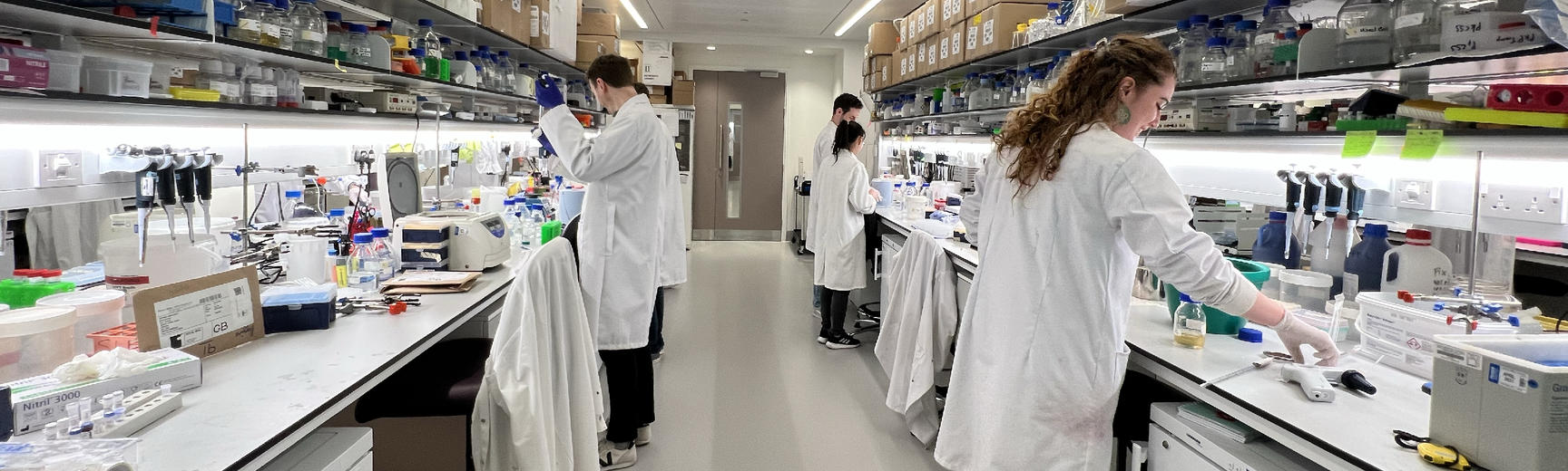
Shared research facilities are at the heart of Kavli INsD. We house state-of-the-art facilities for cryo-EM, super-resolution microscopy, mass photometry and mass spectrometry.
In addition to being open for use by all Kavli members, the facilities can be accessed by researchers from outside our institute or outside the University of Oxford (see below for links to more information on access).

Cryo-electron microscopy (cryo-EM) is one the most powerful techniques for studying the structure and architecture of biological systems. It can be used to study molecules – from individual proteins through to large molecular assemblies and designed nano-structures. It can also be used to probe the structures of cells, revealing their architecture and organisation. With the power to provide detailed insight at a nano-level, this technique is valuable for much of what we do in the Kavli INsD.
The Central Oxford Structural Molecular Imaging Centre (COSMIC) cryo-EM facility is housed in our building. It contains a humidity-controlled ‘prep’ room, three electron microscopes and a computational cluster for data processing. A focused ion beam scanning electron microscope allows preparation of thin layers of cellular material for imaging. An experienced team of facility managers provide training and support, offering world-class cryo-EM access.
For details on the facility and how to access it, please see the cryo-EM facility website.

The nanoimager is a super-resolution fluorescence microscope developed for use in non-specialist laboratories as a benchtop imaging instrument. The instrument offers temperature control, focus correction and multiple illumination modes: epifluorescence/widefield, total internal reflection (TIRF) and HILO (a technique that enables imaging away from the coverslip surface with a high signal-to-noise ratio). It enables users to carry out STORM and PALM super-resolution imaging, as well as single-particle tracking and single-molecule FRET.
For access details, please see the Micron Oxford facility website.

Mass photometry, based on the principles of interferometric scattering microscopy, measures the mass of single molecules with light. The technology has numerous applications, including the study of antibody-antigen interactions, viral assembly mechanisms and receptor-ligand interactions, and the characterisation of samples prior to further analysis (e.g. by cryo-EM).
For further information please see the mass photometry facility website or contact Professor Philipp Kukura.

Complementing the proteomics service in the Biochemistry Department, the Kavli INsD houses a wide range of mass spectrometers dedicated for collaborative projects in nanoscience. Using ‘soft’ ionisation methods and adhering to solution conditions that retain the folded state of the protein, a mass spectrum of a folded protein can be recorded. Such spectra provide insights into complex interactions including protein-protein contacts, lipid or ligand binding as well as antibody conjugation. Coupled with high resolution and highly sensitive detectors, this allows for accurate determination of mass, stoichiometry and, in some cases, small molecule/lipid binding partners.
Further opportunities exist to explore the overall topology of protein complexes in the gas phase via ion mobility measurements and through solution phase labelling techniques, including HDX.
For access, please contact Dr Andrew Dolan, the laboratory manager, or visit the Oxford Mass Spectrometry Centre Instruct-ERIC website (for pan-European, fully funded access).
